Anatomy Of The Upper Chest Area : Upper Chest Wall Anatomy Page 1 Line 17qq Com - It describes the theatre of events.. Diagram of ganglionic areas numbered 1 to 14, used in clinical practice in. Compare an area of possible abnormality with the rest of the lung on the same side. Enlargement will result in bulging of the. Paschalides medical publications, 2004, with permission. • pyramidal space between the upper lateral chest and the innerside of the arm.
Anatomy of peritoneum and mesentery. Paschalides medical publications, 2004, with permission. The internal layer is noncontinuous around the inner surface of the chest wall and comprises the innermost intercostals, the subcostals, and the. It describes the theatre of events. Anatomy of the chest area.
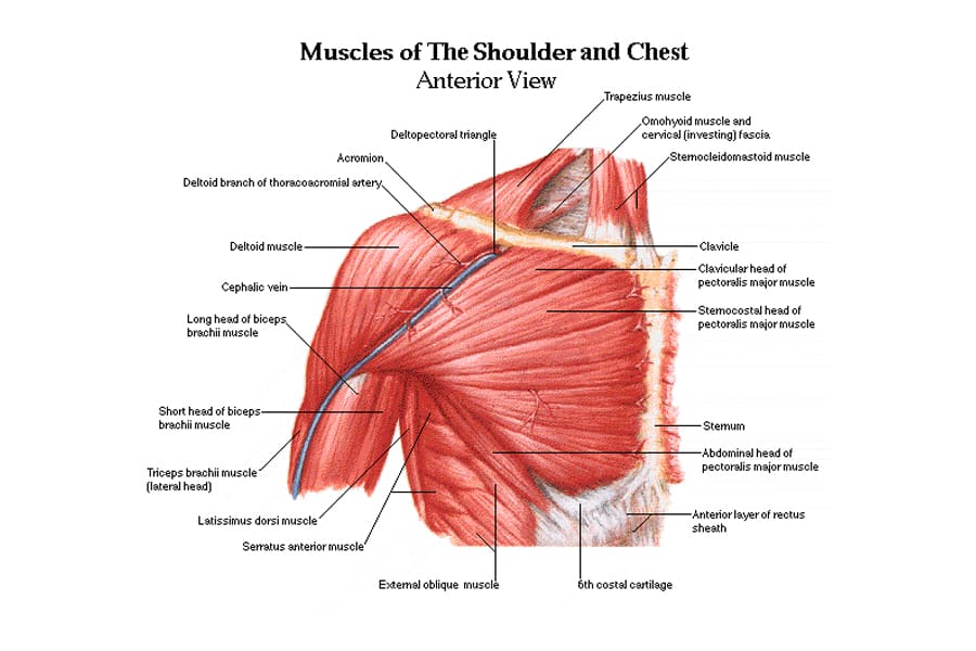
Thoracic Spine Anatomy And Upper Back Pain from embed.widencdn.net The clavicles are attached to the upper lateral part of the manubrium by the sternoclavicular joint. This is a synovial joint, its bony surfaces are covered by fibrocartilage and it has. Anatomy of the chest & abdomen. Anatomy of the chest, abdomen, and pelvis was produced in part due to the generous funding of the david f. • acromion • clavicle • deltoid ( im injections) • humerus axilla(armpit). Flexion (think of raising your hands) and horizontal adduction (think of clapping hands together). Human anatomy for muscle, reproductive, and skeleton. Swensen fund for innovation in teaching.
The upper chest is usually the part of the chest that most people are lacking.
Atlas of anatomy of the human body: Anatomy is to physiology as geography is to history: Surface anatomy of anterior chest wall, spiral ct of thoracic inlet and surface anatomy of posterior chest wall. Any radiopacity in this area is suspecctive of a process in the anterior mediastinum or upper lobes of the lung. Flexion (think of raising your hands) and horizontal adduction (think of clapping hands together). An important palpable feature on the anterior chest wall. Anatomy of the chest & abdomen. Swensen fund for innovation in teaching. Thanks for reading my anatomical guide to training! Enlargement will result in bulging of the. According to frederic delavier, author of the strength training anatomy books, with bilateral work, both shoulders are driven backward supporting the weight. Anatomy is to physiology as geography is to history: We're looking at the anatomy of an upper endoscopy.
The embryologic and anatomic basis of modern surgery. Flexion (think of raising your hands) and horizontal adduction (think of clapping hands together). The opening of the upper chest and thorax. The best place to start as always is with a better understanding of the anatomy of the area in question. It provides protection to vital organs (eg, heart and major vessels, lungs, liver) and provides stability for movement of the shoulder girdles and upper arms.
10 Best Shoulder Exercises For Men Man Of Many from manofmany.com An important palpable feature on the anterior chest wall. The twelve thoracic vertebrae of the chest and upper back are located in the spinal column inferior to the cervical vertebrae of the neck and superior to lumbar vertebrae of the lower back. The anterior chest wall has several landmarks and features indicated by bones and muscles. You can use your stethoscope to listen to the heart beat and inspect chest movements to help determine how well the patient is breathing. It describes the theatre of events. Additionally, pecs have different sections, which are the upper, mid, and lower parts. Chest auscultation requires the chest and back to be exposed, so measures should be taken to this technique allows you to compare one side of the chest with the other in a systematic manner and starting with the upper lobe move to the middle lobe, and finally the lower lobe at the bottom (ferns. The best upper chest workout will.
Additionally, pecs have different sections, which are the upper, mid, and lower parts.
The superior vena cava (svc) is seen in the right paratracheal area, typically representing the right superior mediastinal contour. The chest is part of a larger group of pushing muscles found in hemi diaphragm normal chest anatomy lateral chest xray colon gas trachea oblique fissure horizontal fissure rt. Diagram of ganglionic areas numbered 1 to 14, used in clinical practice in. Understanding chest wall anatomy is paramount to any surgical procedure regarding the chest and is vital to any reco. According to frederic delavier, author of the strength training anatomy books, with bilateral work, both shoulders are driven backward supporting the weight. The anterior of the chest is a main area for physical examination. The lungs are assessed and described by dividing them into upper, middle and lower zones. Enlargement will result in bulging of the. We're looking at the anatomy of an upper endoscopy. The embryologic and anatomic basis of modern surgery. Atlas of anatomy of the human body: Anatomy is to physiology as geography is to history: It describes the theatre of events.
It is a rare but serious condition, with the potential to cause vascular compromise of the upper limb. Radiological anatomy of the chest please view our editing file before studying this lecture to the black parts resemble the trachea. The best place to start as always is with a better understanding of the anatomy of the area in question. The embryologic and anatomic basis of modern surgery. An important palpable feature on the anterior chest wall.
Upper Chest Anatomy Page 1 Line 17qq Com from img.17qq.com This is a synovial joint, its bony surfaces are covered by fibrocartilage and it has. The upper posterior border of the heart is formed by the left atrium. Related posts of anatomy of the chest area. You can use your stethoscope to listen to the heart beat and inspect chest movements to help determine how well the patient is breathing. Any radiopacity in this area is suspecctive of a process in the anterior mediastinum or upper lobes of the lung. The chest anatomy includes the pectoralis major, pectoralis minor and the serratus anterior. Anatomy is to physiology as geography is to history: It is not uncommon for someone to have an underdeveloped upper or lower chest or maybe even wish they had better definition in the inner or outer chest region.
Atlas of anatomy of the human body:
All about the chest muscles function of the chest muscles. It describes the theatre of events. Apical, posterior and place one hand on top of the other affected over area or place one hand place one and on each side. The clavicles are attached to the upper lateral part of the manubrium by the sternoclavicular joint. Seen clearly crossing the upper part of each lung field. Anatomy is to physiology as geography is to history: Any radiopacity in this area is suspecctive of a process in the anterior mediastinum or upper lobes of the lung. Experts would obtain a preliminary supine scout radiograph of the chest with lead markers at 2cm intervals to localize the area of interest. Paschalides medical publications, 2004, with permission. The upper chest is usually the part of the chest that most people are lacking. • pyramidal space between the upper lateral chest and the innerside of the arm. Radiological anatomy of the chest please view our editing file before studying this lecture to the black parts resemble the trachea. Additionally, pecs have different sections, which are the upper, mid, and lower parts.


Posting Komentar
0 Komentar